These research results were gained through a joint effort with a group led by Simon Cherry, a distinguished professor at UC Davis School of Medicine in the US, a group led by Professor Yoichi Tamagawa at University of Fukui in Japan, and with Professor Tomoyuki Hasegawa at Kitasato University in Japan. The major results were published on Thursday, October 14, 2021 in the electronic edition of “Nature Photonics”, a British scientific journal.
Research work background
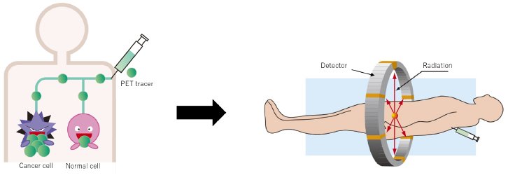
In making a diagnosis with a PET device, a PET tracer that easily accumulates in cancer cells is administered to the patient, and the radiation emitted from sections where the tracer accumulates is then detected from various angles by detectors laid out in a ring shape to discover the cancer cells. Currently, the best system time resolution that shows the detection accuracy of the position where the radiation is emitted is approximately 200 picoseconds (a picosecond or ps, equals one trillionth of a second). This accuracy allows the measurement of the radiation emission position within an error of approximately 3 centimeters. However, to obtain even higher accuracy, the target position is measured within an error of approximately 0.4 centimeters by obtaining a huge amount of data and reconstructing the images. To attain high accuracy equal to, or better than conventional imaging systems by using a pair of detectors and without doing image reconstruction, a time resolution of approximately 30 picoseconds is required. So at Hamamatsu Photonics, in addition to moving ahead with developing detectors with higher time resolution utilizing our photomultiplier tubes, we have also performed experiments with a PET tracer synthesized within the company’s own premises.
Overview of research results
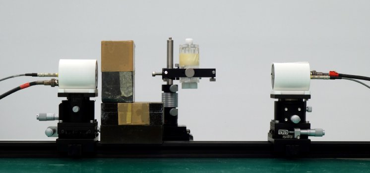
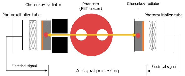
Ordinary PET devices work by using scintillators to convert the radiation from within the body into scintillation light 1) and detecting it. A higher time resolution can be achieved by utilizing Cherenkov light 2) that has a higher response speed to radiation than scintillation light, but Cherenkov light has the problem of low light emissions. To deal with this issue, we developed a new MCP-PMT (microchannel plate – photomultiplier tube) with high sensitivity and high time resolution that also incorporates a Cherenkov radiator for efficiently converting radiation into Cherenkov light. Using this new MCP-PMT allows for the detection of very low Cherenkov light with high sensitivity. We also succeeded in getting even higher time resolution by using a newly developed signal processing technique that makes use of AI (artificial intelligence). In this way, for the first time in the world we achieved a time resolution of approximately 30 picoseconds and our experiments using a PET tracer succeeded in obtaining high-accuracy medical diagnostic imaging from a pair of detectors, without having to perform image reconstruction processing.
Applying the results from these research efforts will lead us to achieve a completely innovative, simple and compact measurement device that uses a pair of detectors capable of medical imaging with high accuracy equal to or better than conventional radiation imaging systems such as PET and CT that need to acquire a huge amount of data by using a ring of radiation detectors. In this way, in addition to boosting inspection efficiency and the speed of diagnosing diseased tissues or organs such as from cancer, the radiation exposure dose can be reduced to alleviate the burden on the patient and medical staff.
We will continue our R&D work to achieve even better time resolution and practical product applications.
1) Scintillation light: Light emitted when radiation reacts with a scintillator
2) Cherenkov light: Light emitted when a charged particle passes through a material at speeds faster than light
For further information contact us on email: europe@hamamatsu.de or visit our website: www.hamamatsu.com


