The method is based on the atomic force microscope (AFM), an instrument invented by an IBM Nobel Laureate 20 years ago that performs nanoscale operations using a tiny cantilever with a cone-shaped tip at its end. When an electrical field is applied to the tip, molecules will slide up or down its surface at characteristic speeds. By modifying the tip's surface and varying the strength and duration of the electric field, different molecular species can be separated from each other within a few milliseconds, more than 1,000 times faster than today's methods.
"Our initial tests used fragments of DNA – one with five base pairs, another with 16," said H. Kumar Wickramasinghe, IBM Fellow and co-developer of several different types of AFMs. "An electric field propelled these molecules down the 11.2-micron length of the AFM tip in 5 and 15 milliseconds, respectively. We controlled the passage of as few as 10 molecules, which indicates that this approach should be very useful for analyzing very small biological samples and in writing extremely small features."
The new method is described in a technical report published in the May 1, 2006, online edition of professional journal Applied Physics Letters: "Ultrafast molecule sorting and delivery by atomic force microscopy" by Kerem Unal, Jane Frommer and Wickramasinghe, who are researchers at IBM's Almaden Research Center in San Jose, California.
The IBM scientists anticipate that their new method could someday be used in at least two very different applications. Since it is a highly miniaturized version of "electrophoresis," a standard procedure for using electric fields to separate biological molecules, the new method may speed up a wide variety of molecular analysis and genetic applications – from DNA fingerprinting to routine blood tests.
"This very clever use of an atomic force microscope has the potential to provide ultra-fast and ultra-sensitive molecular separations," said Dr. David Garfin, a Berkeley, Calif., consultant and president of the American Electrophoresis Society. "Today, electrophoresis separations in gels or capillary tubes typically take minutes to hours. It would be a tremendous improvement if this early-stage technology can be developed to deliver over a wide range of molecular sizes the precise sub-second separations that the IBM scientists have demonstrated."
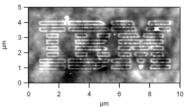
It also has potential for delivering molecules onto a surface with great precision, which may be useful in creating future molecular electronic circuits or lithography features for more conventional nanoelectronics. To demonstrate the technique's initial deposition prowess, the scientists used a single tip to write an outline of IBM's classic 8-bar corporate logo in 59-79-nanometer-wide lines composed of 5-base-pair fragments of DNA. The logo was so small that more than 300 of them would fit on the cross section of a human hair.
IBM's new technique has a number of advantages over existing AFM molecule deposition methods, such as "dip-pen lithography," which place dissolved molecules on a surface by a process similar to a fountain pen writing ink on paper.
"Our new method acts more like an inkjet printer than a fountain pen," Wickramasinghe said. "For example, we write only when the electric field in applied, not continuously while in contact with the surface. We can also control the deposition rate by varying the electric field strength, and our new technique is much less sensitive to the chemical properties of the molecules or the writing surface."
IBM is exploring options for commercializing this invention.
IBM Research has a distinguished history in developing microscopes for nanoscale imaging and science. Gerd Binnig and Heinrich Rohrer of IBM's Zurich Research Laboratory received the 1986 Nobel Prize in Physics for their invention of the scanning tunneling microscope, which can image individual atoms on electrically conducting surfaces. Also in 1986, Binnig invented the AFM, which measured the various forces between a cantilever tip and features on non-conducting surfaces. Scientists at IBM and elsewhere modified and extended the AFM design to image surface forces such as magnetism, friction and electrostatic attraction with nanometer resolution. In 2004, IBM scientists detected the spin of a single electron using an AFM variant known as a magnetic resonance force microscope that they had designed and built.

