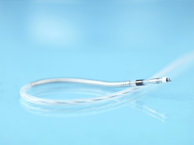
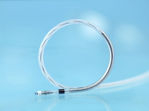
“The average person’s heart beats more than 100,000 times a day, so you can imagine that ICD leads undergo a substantial amount of movement and bending with every contraction. For this reason, leads must be able to endure a lot of stress,” said Dr. Jan Schmidt, Clinic for Cardiology, Angiology und Pneumology at the University Hospital in Düsseldorf.
Plexa ProMRI is the culmination of eight years of research and development to continuously further enhance the quality of ICD leads. The new lead offers enhanced resilience through a performance-driven helical design that is resistant to bending stress. The helical design concept was introduced to ICD leads by BIOTRONIK in 2013 with the DF4 connector. With the launch of Plexa ProMRI, for the first time the benefits of this design have been brought to the intracardiac region, between the right atrium and ventricle at the tricuspid valve—one of the most demanding areas for ICD leads.
“Taking the low impedance conduction of ICD leads and the high durability of pacemaker leads, Plexa ProMRI combines the best of both worlds to offer an ICD lead that can face high levels of stress in the areas it is needed the most,” said Dr. Schmidt, one of the first physicians to implant the Plexa ProMRI lead.
BIOTRONIK’s ICD leads are already recognized for long-term safety and reliability, as demonstrated in the GALAXY and CELESTIAL studies.1 “Longevity is extremely important for patient care when it comes to leads for implantable cardiac devices,” said Manuel Ortega, Senior Vice President at BIOTRONIK. “For this reason, we have dedicated several years to developing an ICD lead that seeks to surpass the standards and reinforce patient safety.”
About Plexa ProMRI
Plexa ProMRI allows patients to undergo MRI scans when used in combination with the relevant MR conditional devices. The new lead is also designed to facilitate handling during implantation with modified accessories that simplify the procedure. Moreover, it supports DX technology for ICDs and now also CRT-Ds, enabling atrial sensing in patients without an atrial lead.
Reference
1 Good ED et al. Journal of Cardiovascular Electrophysiology. 2016, 27(6).


