The connected doctor serves patients even outside the practice
Mobile, digital, connected, these are the trends in skin cancer detection. Therefore, more and more doctors use a digital handheld dermatoscope in addition to their local imaging system. handyscope converts an iPhone into such a device. It has been well established among doctors, enabling them to take digital mole pictures independently of a computer - in clinic and practice, on the way or at home visits.
Now there are new possibilities to work with these mobile images. They can be uploaded into a secure private web space on the cloud-like internet platform FotoFinder Hub. From there it is possible to request a second opinion on suspicious moles. A team of international skin cancer experts reviews the pictures and sends a rating within minutes. Furthermore, doctors can establish private networks to exchange information about suspicious cases. These possibilities through eDermoscopy give more patients access to latest diagnostic standards. handyscope pictures can now also be integrated in local FotoFinder systems; pictures are transferred via WiFi and saved in the central database .
handyscope is presented at the AppCircus on Medica Health IT Forum, hall 15, booth 15A03 on November 16, 2012.
Skin analysis with Body Mapping
The earlier skin cancer is detected, the sooner treatment begins. The latest method for early detection comprises a regular monitoring of the entire skin and each individual mole.
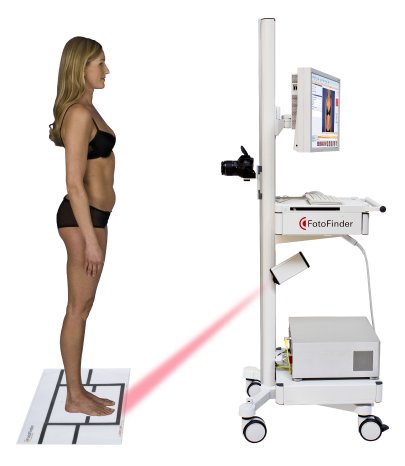
The entire skin surface is documented in a standardized way with FotoFinder bodystudio. New SLR technology delivers photos with 18 megapixels. The entire imaging process is controlled by the software and guides the user. In this way, four pictures are taken from each side of the body and stored immediately. The result is a "digital skin map" where the doctor easily identifies suspicious moles. In addition, these moles are photographed dermoscopically with a magnification of 20X to 70X.
At regular check-ups there is no need for research in paper files, because the entire skin status is saved in the software. Follow up images of the full body photographs as well as those of each mole show changes at a glance. The expert software Bodyscan pro compares initial and follow up images and shows new or altered moles whereas the Moleanalyzer analyzes the microscopic structure of single lesions and gives a score for malignancy.
The combination of Total Body Photography and digital dermoscopy helps dermatologists, not only if they examine patients with multiple moles. It gives patients security that nothing is overseen and helps to identify skin cancer at an early stage. "Nowadays physicians have less and less time for their patients, on the other hand the skin is a large organ which demands a fine examination. This contradiction is solved with the timesaving, but accurate Body Mapping in combination with dermoscopy", explains Andreas Mayer, CEO of FotoFinder Systems.

