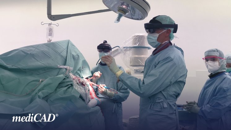
The surgical plan was exported both as a 3D model and a detailed planning report with dimensions, which were transmitted to the two HoloLenses used at the beginning of the procedure. During the surgery, both surgeons had access to the same hologram of the intended outcome, using the 3D model to visualize and comprehend the positioning of humeral head fragments. A critical aspect was the correct placement of the K-wire and baseplate on the glenoid to achieve the planned implant position. Thanks to the 3D model of the scapula, the surgeons were able to understand the glenoid's structure, even with soft tissues obstructing the patient's anatomy. The Friedmann line displayed on the hologram served as an additional reference point to achieve an optimal result.
The simultaneous transmission of the surgeons' perspective through video communication allowed the rest of the surgical team to follow the current status of the procedure and prepare for the next steps promptly. Ultimately Lastly, the operation was successfully completed, thanks to the intraoperative support provided by the latest mediCAD® software. Users were delighted with its user-friendliness and intuitive interface.
mediCAD would like to express gratitude to Dr. Rieske and his team for the opportunity to collaborate and utilize this fascinating new technology in the operating room! By employing mediCAD 3D software, surgeons were able to create more precise plans and access crucial information during the surgery, leading to a successful outcome. This advancement in medical imaging and planning holds promising prospects for the future of orthopedic surgery.


