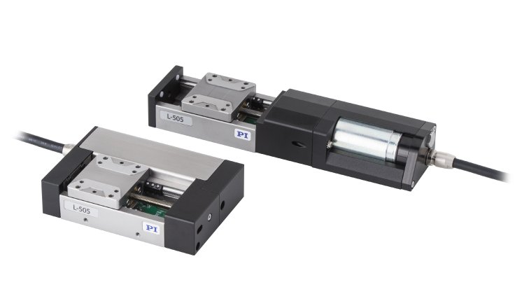
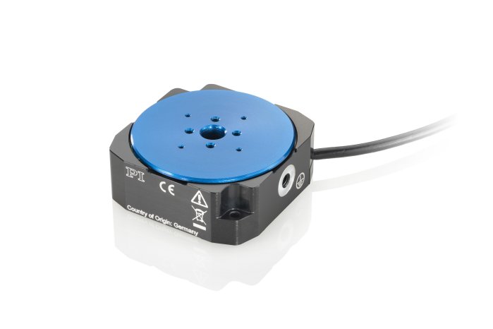
"We aim to democratize high-end light microscopy, bringing it to campuses and labs for free," explains Professor Jan Huisken, co-inventor of light sheet microscopy and Director of the Medical Engineering Department at the Morgridge Institute for Research. The project, which is supported by several sponsors, turns the idea of central research facilities upside down and makes travelling with sensitive samples obsolete.
Up to now, two complete systems have been set up; for the coming months and years, Huisken and his team are planning to build several dozen Flamingo-type microscopes. The name derives from the shape of the basic version of the microscope, which reminds us of the bird standing on one leg.
Light sheet microscopy
In Light Sheet Fluorescence Microscopy (LSFM), illumination and detection are two separate optical systems that are oriented perpendicular to each other. A laser beam is focused to form a light sheet that illuminates a thin plane within the sample. The fluorescent light that is emitted in this plane is captured and detected by an objective lens. "LSFM is a very powerful and flexible platform for gentle in vivo imaging, offering low phototoxicity and fast image acquisition," Jan Huisken summarizes the advantages of the technology. Because of the gentle "treatment" of the sample, LSFM is predestined for the examination of living organisms.
For more details also visit the respective blogpost at https://www.physikinstrumente.com/en/pi-blog/.
About the Huisken Lab at the Morgridge Institute for Research
The Huisken Lab is creating tailor-made optical microscopes to non-invasively explore morphogenesis and function in living organisms. The Morgridge Institute for Research is an independent biomedical institute exploring uncharted scientific territory to discover tomorrow’s cures.


