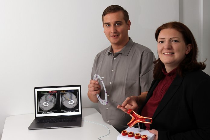
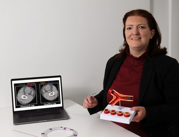
Too little exercise, unhealthy food, smoking - these factors promote arteriosclerosis. In industrialized countries, the disease is responsible for half of all deaths. “It is usually only discovered when it has already advanced,” says Christina Gillmann from the Technische Universität Kaiserslautern (TUK). “On CT images, physicians can only recognize these deposits, if thicker layers are already present on the vessel walls.” The only therapy that can then be considered at this point is surgery.
However, if arteriosclerosis is detected at an early stage, it can be treated with a healthy diet and exercise. That's what the computer scientists around Gillmann are working on. They are developing a computer programme to help doctors make an early diagnosis. In this method, already existing CT images are used. This X-ray method provides doctors with patient images in layers, which are usually shown in grayscale. “The resolution of these images is not very high,” says the computer scientist. “To detect arteriosclerosis at an early stage, the data must be processed in a different way.”
Gillmann and her team screen the CT images for additional information with their software. For example, they are able to accurately represent the arterial bifurcations and better classify and locate the progress of the disease. In addition, the computer scientists analyse different types of catheters that can be used during the operations. In this way, the best possible solution can be found for the individual patient and the risk of potential complications during and after the surgery can be reduced. The researchers at the TUK are developing their system in collaboration with doctors from Dayton in the USA under Professor Dr Thomas Wischgoll and from Colombia under Professor Dr José Tiberio Hernández Peñaloza.
However, a few more years of development work will be needed before the system can be used in hospitals. The method will also be of interest to industrial companies. They could, for example, use the technology to screen their products more specifically in order to detect possible damaged areas.
At Medica, the scientists will present their method at the research stand of Rhineland-Palatinate.
Gillmann is researching in the field of “Computer Graphics and Human Computer Interaction” under Professor Dr Hans Hagen. For about two decades now, the research group has been working on processing medical imaging data in such a way that they can be used easily and reliable in everyday clinical practice. For example, they have succeeded in using their methods to separate tumours from healthy tissue more clearly in images. In their projects, the computer scientists work closely with various partners, including the University Hospital Leipzig and the Premier Health Hospital in Ohio.


